
· Symptoms of acute infection of elephantiasis are:
1) Fever
2) Pain in lymph nodes
·
3) swollen lymph nodes
·
4) Thickening skin
24 June 2005 - Simple antibiotics may offer a cure for the previously “incurable” tropical  disease, elephantitis, according to a study published in the May issue of The Lancet. Parasitologists from the University of Bonn and in Hamburg, Liverpool and Tanzania found that the common antibiotic doxycyclin kills the threadworms that cause elephantitis. Current drugs for elephantitis can kill the worm larvae, or micro-filariae, but have no affect on the adult worms. In addition, these drugs can have serious side-effects.“Due to the long lifespan of the wuchereria worms, therapy lasts several years, during which time the symptoms continue to persist,” said University of Bonn professor Achim Hörauf.Hörauf and his colleagues found that adult wuchereria worms can be killed indirectly by killing essential bacteria that live inside the worms. After 14 months of treatment with the antibiotic doxycyclin, only 20% of elephantitis patients still had living adult worms, compared to 89% of a placebo group.Doxycyclin is already used to treat respiratory and gastro-intestinal infections, and it is relatively cheap. Another advantage is that this antibiotic only has minor side-effects.“The importance of these findings for therapy should not be underestimated,” Hörauf emphasises. “The mature worms are after all responsible for such symptoms of the disease as the extreme swelling of the limbs. In the past there was no effective and reliable method of combating them.”
The Journal of Young Investigators. New Treatment For Elephantitis: Antibiotics [Data file]. Retrieved from:
http://www.jyi.org/news/nb.php?id=361
The drug of choice in treating lymphatic filariasis is diethylcarbamazine (DEC). The trade name in the United States is Hetrazan.
The treatment schedule is typically 2 mg/kg per day, three times a day, for three weeks. The drug is taken in tablet form.
DEC kills the microfilariae quickly and injures or kills the adult worms slowly, if at all. If all the adult worms are not killed, remaining paired males and females may continue to produce more larvae. Therefore, several courses of DEC treatment over a long time period may be necessary to rid the individual of the parasites.
DEC has been shown to reduce the size of enlarged lymph nodes and, when taken long-term, to reduce elephantiasis. In India, DEC has been given in the form of a medicated salt, which helps prevent spread of the disease.
The side effects of DEC almost all are due to the body's natural allergic reactions to the dying parasites rather than to the DEC itself. For this reason, DEC must be given carefully to reduce the danger to the individual. Side effects may include fever, chills, headache, dizziness, nausea and vomiting, itching, and joint pain. These side effects usually occur within the first few days of treatment. These side effects usually subside as the individual continues taking the drug.
There is an alternate treatment plan for the use of DEC. This plan is designed to kill the parasites slowly (to reduce allergic reactions to the dead microfilariae and dying adult worms within the body). Lower doses of DEC are taken for the first few days, followed by the higher dose of 2 mg/kg per day for the remaining three weeks. In addition, steroids may be prescribed to prevent the individual's body from reacting severely to the dead worms.
Another drug used is Ivermectin. Early research studies of Ivermectin show that it is excellent in killing microfilariae, but the effects of this drug on the adult worms are still being investigated. It is probable that patients will need to continue using DEC to kill the adult worms. Mild side effects of Ivermectin include headache, fever, and myalgia.
Other means of managing lymphatic filariasis are pressure bandages to wrap the swollen limb and elastic stockings to help reduce the pressure. Exercising and elevating a bandaged limb also can help reduce its size.
Surgery can be performed to reduce elephantiasis by removing excess fatty and fibrous tissue, draining the swelled area, and removing the dead worms.
The Free Dictionary by Farlex. Treatment [Data File]
Retrieved from:
http://medical-dictionary.thefreedictionary.com/Elephantitis
 Acidic Soap Step 2: Phanta Soaking
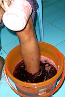
Step 3: Yoga Step 4: Massage


Step 5: Compression

INTEGRATED TREATMENT OF LYPHODEMA/LYMPHATIC FILARIASIS
A low cost treatment protocol was outlined by Vaqas & Ryan (2002) to replace the high cost systems of management, termed complex decongestive physiotherapy (CDP), originating in Europe. Their low cost management technique for resource poor settings suggests improving the lymphatic flow by accentuation of physiologic mechanisms of lymph drainage and good skin hygiene. Based on their outline, initially a case study was conducted at the Institute of Applied Dermatology on a 60 years old female patient with 36 years duration of lower limb lymphoedema. We undertook this larger study with an aim to develop an effective and alternative method of treatment to achieve the morbidity reduction at low cost in filarial lymphoedema patients. In Ayurveda, filarial manifestations are described as Shleepada, from the Sanskrit terms Shila = stone and Paada = foot, i.e. stony appearance of the foot. Now we have succefuly treated over 350 patients of lower limb lymphodema and published finding in World’s leading journals on lymphology
TREATMENT METHODOLOGY:The treatment included an initial 14 days of hospitalization followed by 6 months of treatment at home.
Patients are admitted in a KPO (Knowledge process outsourcing) hospital, of Institute of Applied Dermatology. Baseline data are recorded on indicators of improvement and the status of the skin was documented with photographs. The entire team attending the patient makes the baseline assessment of the patient. The Ayurvedic doctor selects the ayurvedic herbal medicines used for the patient after discussions with the dermatologists in the team. In the case of multiple nodules on the limb, the homeopathy doctor selects their drugs after discussions with the other members of the team. Before the onset of treatment, each patient’s management strategy and details are discussed in the team. In case of difficult cases details are being e-mailed to Prof Terence J Ryan, Emeritus professor, department of dermatology, Oxford Medical School and member of the lymphatic filariasis-working group on clinical management trials under GAELF for his agreement & advice.
DIFFERENT STAGES OF TREATMENT :
Skin care measures: this includes treating the skin with ayurvedic skin tonic called phanta. Yoga Pretreatment: Following the phanta soaking, a series of yoga exercises are performed. Pre and post treatment yoga exercises are taught to the patient by a yoga therapist with special attention to breathing co-ordination. Pre and post treatment yoga exercises are performed as explained in English translations of classical yoga texts. A yoga therapist who spends 2-3 hours a day with the patient, coaches yoga exercises. A CD-rom containing the complete details of yoga exercises is provided to the patients. Manual lymph drainage of central lymph nodes, a mandatory pre treatment procedure as part of Foldi’s complex decongestive physiotherapy practiced in Germany is not performed in our patients. Instead, series of Yoga exercises done. Indian Manual Lymph drainage (IMLD): Immediately following pre treatment yoga patients are subjected to IMLD. Compression Bandaging: 20 minutes after IMLD compression bandages are applied using the products of an Indian manufacturer. Post treatment yoga: Post treatment yoga is done wearing the compression bandage and on an empty stomach
METHODOLOGY:
Pretreatment counseling is given using the power point presentation with evidence for the reduction in limb size and improvement in quality of life scores to all the patients as routine. Once convinced the patients are requested to fix a suitable date for their treatment.
During the two weeks stay in the hospital the patient and a family member is trained to perform all these activities so as to carry them out at home. Handouts containing the details are given in their local language. Comprehensive education about the importance of each component of ongoing therapy is given to every patient. Patients are strictly advised to stop monthly penicillin injections or antibiotics, if they take as a preventive measure for inflammatory episodes. The diuretic Frusemide is discontinued unless the patient has signs of heart failure
CONTINUATION OF THERAPY: Patients are asked to perform all these procedures, everyday for six months in the sequence as trained during hospitalization. In addition they are advised to take two oral medications of Ayurveda described for lymphoedema, as recommended by ayurvedic formulary of India. DIET: Patients are advised to observe certain diet restrictions while on oral medications. They are advised to avoid cold water & cool drinks, milk and milk products, black gram, horse gram, cashew nut, ground nut, tamarind, jaggery, sesame and brinjal and to be strictly vegetarian in diet.
FOLLOW UP:Patients are scheduled for follow up at 4 weeks, 12 weeks and 24 weeks following the discharge. During each follow up, the patients shall demonstrate procedures; they are doing at home including all the exercises of yoga in order to perfect the procedures.
This treatment is to be carried out at home for life long
Three national seminars held during 2005 2007& 2008 supported by Indian Council for Medical Research , Department of Science & Technology, New Delhi ,department of AYUSH New Delhi & Kerala State Council for Science, Technology and Environment, Thiruvananthapuram has approved this new treatment and recommended that the new treatment developed as through international collaboration may be adopted for the national program

Before After
Institute of Apllied Dermatology. Integrated Treatment for filariasis and lymphoedema [data file]. Retrieved from:
http://www.indiandermatology.org/our-services/integratedfilariasistreatment/index_html

